Maximising our efforts during cardiac resuscitation, means maximising our chest compressions to achieve the best cardiac output possible. The reality is that we don't know the best point of compression, to achieve output and surely it cannot be the same for everyone.
Recommendations include the lower half of the sternum, or the internipple line; all based on external landmarks rather than true anatomy. There are indications in the literature that the ideal area of maximal compression(iAMC) is over the maximal diameter of the left ventricle (JTrauma Acute Care Surg 2018;85(2):303–10). The study did not aim to establish that the iAMC is physiologically or clinically the optimal compression location.
This study used transthoracic echocardiography to see if the guideline-recommended chest-compression position over the lower half of the sternum aligned with the maximal diameter of the left ventricle, or with with the LVOT?
THE STUDY
Martinez L, et al. Where should we compress? Ultrasound identification of the ideal area of maximal compression for CPR Resuscitation. 2026;226:111220.
Why this matters
High-quality CPR is usually defined by compression rate, depth, recoil and minimising interruptions. Much less attention has been paid to exactly what cardiac structure lies beneath the rescuer’s hands.
Current landmark-based CPR may produce two competing effects:
If sternal compressions preferentially compress the left ventricular outflow tract (LVOT), they could obstruct ventricular ejection and reduce forward flow—even when the compression rate and depth are technically perfect.
Previous intra-arrest transoesophageal echocardiography studies have shown frequent LVOT compression and have suggested that moving compressions laterally may improve blood pressure and end-tidal CO₂. However, the anatomical relationship between external landmarks, the LVOT and the left ventricle remains incompletely characterised.
What They Did
This was a prospective, single-centre observational anatomical study conducted in an academic emergency department.
A convenience sample of 152 clinically stable adults (>18yo), comprising emergency department patients and healthy volunteers, enrolled between July 2024 and December 2025.
Key characteristics:
| Characteristic |
Result |
| Median age |
62.5 years |
| Male |
67.1% |
| Median weight |
82 kg |
| Median height |
173 cm |
| Previous intrathoracic surgery |
17.8% |
| Current pericardial effusion |
2.6% |
Intervention and measurements
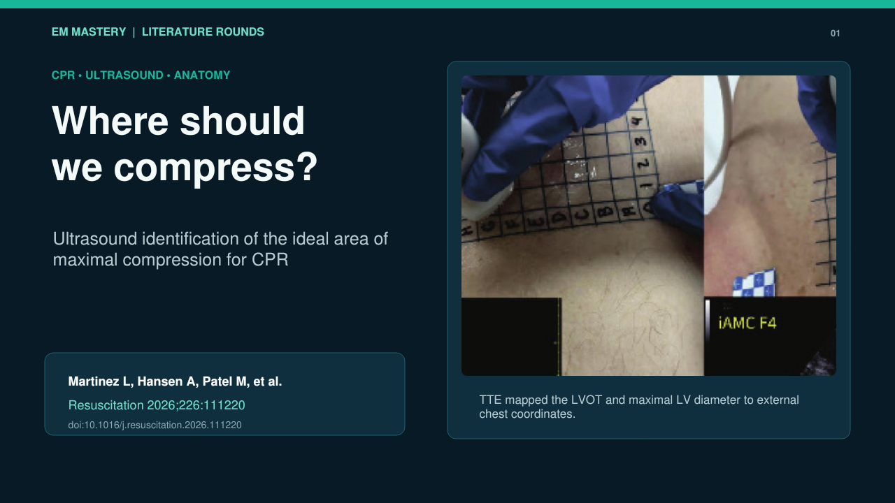
Investigators used transthoracic echocardiography to map cardiac structures onto a transparent 1-cm grid placed over the anterior chest.
The grid was referenced to the xiphoid process and sternum as the image reproduced below shows.
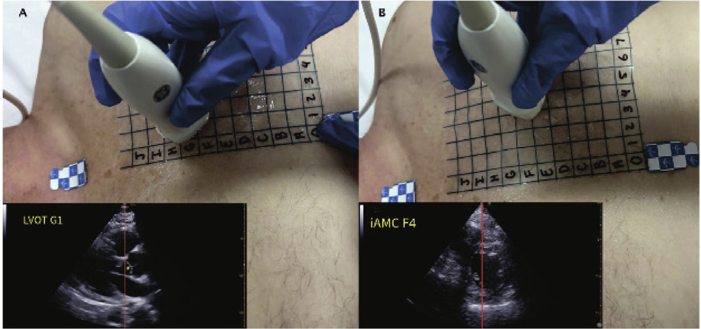
In the above illustration (A) The parasternal long-axis view of the heart (lower left) is aligned with the red midline, and the target structure – in this example, the LVOT (yellow asterisk) – is centered and recorded at its corresponding grid location (G1).
(B) This is the parasternal short-axis view at the level of the papillary muscles to identifies the ideal area of maximal compression (iAMC)
Adhesive stickers mark the sternal notch and xiphoid process which were identified with ultrasound.
Two anatomical targets were identified:
-
LVOT: located using the parasternal long-axis view.
-
Ideal area of maximal compression (iAMC): defined as the maximal diameter of the left ventricle at the papillary-muscle level in the parasternal short-axis view.
The external positions of these structures were then compared with the guideline-recommended sternal compression zone.
Primary Outcome
"The primary outcome was characterization of the external chest wall locations of the LVOT and iAMC relative to the guideline-recommended sternal chest compression target."
What They Found
| Anatomical structure |
Median horizontal distance from sternum |
| LVOT |
1.5 cm left, IQR 1–2 cm |
| iAMC |
4 cm left, IQR 3–5 cm |
The LVOT lay within 1 cm of the sternum in 50% of participants.
The iAMC lay within 1 cm of the sternum in only 2.7%.
Heat maps showed that:
Authors’ conclusion
Guideline-recommended sternal compressions may frequently be delivered over the LVOT rather than the maximal diameter of the left ventricle. They do not recommend changing current CPR practice on the basis of this study.
Strengths
-
Prospective data collection using a standardised grid.
-
Reasonably sized cohort for an anatomical mapping study.
-
Direct ultrasound identification of cardiac structures rather than inference from surface landmarks.
-
Included older emergency department patients as well as volunteers.
-
Provides a plausible anatomical explanation for LVOT obstruction observed during intra-arrest TEE.
-
Addresses an important component of CPR quality that has received relatively little attention.
Important limitations
1. This was not a cardiac-arrest study
Participants were stable and supine but were not receiving chest compressions. Cardiac position and thoracic geometry may change during arrest due to chest deformation due to compressions, loss of vascular tone and filling, or gastric distension.
Static anatomy therefore may not represent the structure actually compressed during CPR.
2. No physiological or clinical outcomes
The study was not designed to measure clinical outcomes. The study showed structure location not where compressions generate the best output.
3. The “ideal” target is assumed
The iAMC was defined as the maximal LV diameter at the papillary-muscle level and was assumed to be the “ideal area of maximal compression”. The study therefore cannot tell us whether moving the hands approximately 4 cm to the left would improve or worsen CPR.
4. Selection and generalisability
This was a single-centre convenience sample, with 67% male participants. Patients with inadequate TTE windows were excluded, potentially removing people with obesity, hyperinflated lungs or other anatomy in whom targeting might be most difficult.
5. Operator and measurement limitations
The scans were performed by ultrasound fellowship-trained clinicians, however we are not sure of the reproducibility of the data.
6. Safety remains unknown
The effect of more lateral compressions was not assessed. Risks that could be associated with such a position may result in rib and pulmonary injury, splenic or hepatic injury, depending on position.
Interpretation
This paper asks looks to see the anatomical alignment of chest compressions. However, anatomical alignment does not automatically translate into better haemodynamics or better patient outcomes.
The moderate correlation between LVOT and iAMC positions also shows why a universal instruction such as “move 4 cm left” is unlikely to be anatomically precise for every patient.
The findings support further trials of ultrasound- or TEE-guided compression positioning, but they do not yet support changing routine hand placement.
Take-home message
During conventional CPR, we may often be compressing the LVOT rather than the left ventricle. The use of transthoracic or transoesophageal echocardiography may asist us in determing an optimal compression zone to achieve the best output during CPR. This is currently being done in some emergency departments. If these investigation modalities are not available then we keep compressing over the lower half of the sternum.