Not all ventricular tachycardias have very wide QRS complexes, and not all regular, relatively narrow-complex tachycardias are supraventricular.
Fascicular ventricular tachycardia (FVT), also known as verapamil-sensitive ventricular tachycardiaor Belhassen tachycardia, is not your normal VT. It is not wide. Verapamil is generally contraindicated in undifferentiated wide-complex tachycardia because of the risk of haemodynamic collapse. The exception applies to confidently diagnosed, haemodynamically stable, verapamil-sensitive fascicular VT—not simply any regular tachycardia with an RBBB pattern.
Case
A patient in his40s, presents to the emergency department with sudden-onset palpitations. He feels lethargic, butis haemodynamically stable. He has no significant past medical history.
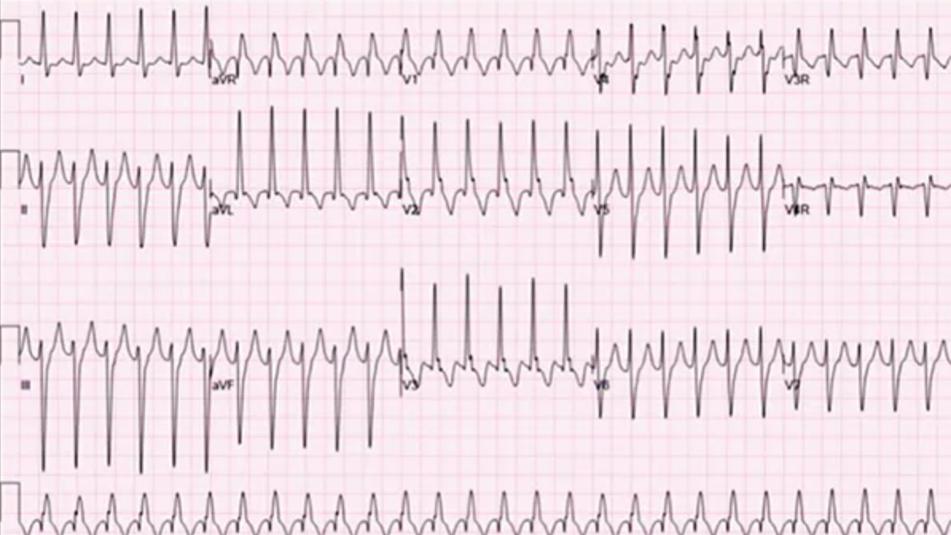
His ECG is shown below. He is treated with escalating doses of Adenosine with no success. Metoprolol has no effect. He is DC cardioverted, however returns to the tachycardia again.
What does the ECG show and what is the diagnosis? Given that this is a review of fascicular VT, we know the answer, however there are other potential diagnoses of regular narrow tachycardia that may fool us.
ECG interpretation
On initial consideration, it looks like an SVT with RBBB, but let's look more closely:
ECG feature
Interpretation
Rhythm
Regular tachycardia
QRS morphology
Right bundle branch block pattern
Frontal axis
Left axis deviation
QRS duration
Relatively narrow for VT, typically under 140 ms
Lead aVR
Positive QRS
Lead V6
Deep S wave, R/S ratio ≤1
Atrial activity
No clear associated P waves
The combination of RBBB morphology and left axis deviation strongly suggests left posterior fascicular VT in the appropriate clinical context. There are also no p waves or AV Dissociation in FVT, a positive QRS in aVR Left axis deviation and R/S ratio < 1 in V6
A key diagnostic pitfall is that the QRS may be only modestly prolonged, making the rhythm resemble SVT with aberrant conduction.
Why can't it be one of the other choices?
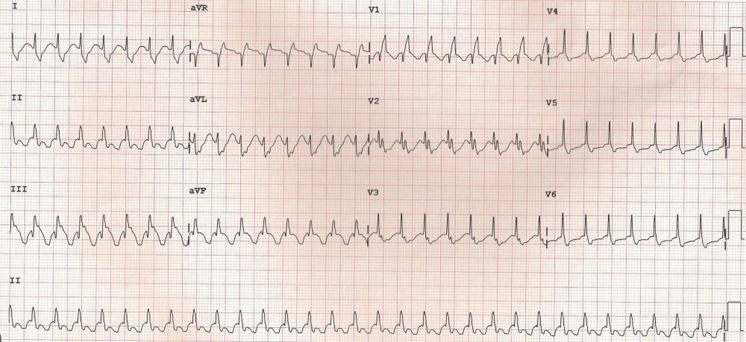
Atrial Fibrillation: It is regular Atrial Flutter: There is no sawtooth pattern SVT + RBBB: There will usually be p waves visible, and a negative QRS in aVR
Below is an example of SVT with RBBB(Johnsonfrancis.org). Compare the ECGs. This is a narrow complex with evidence of RBBB in several leads.
Why is fascicular VT different?
Fascicular VT is an idiopathic ventricular tachycardia that typically occurs in younger patients without structural heart disease.
Unlike most ventricular tachycardias, its re-entry circuit involves specialised Purkinje tissue and a slow-conducting, calcium-dependent pathway.
There are three recognised patterns:
Type
Typical ECG pattern
Left posterior fascicular VT
RBBB with left axis deviation — most common
Left anterior fascicular VT
RBBB with right axis deviation
Upper septal fascicular VT
Often relatively narrow QRS, sometimes near-normal axis
The case represents the left posterior fascicular form.
How todistinguish fascicular VT from SVT with aberrancy?
Feature
Fascicular VT
SVT with RBBB aberrancy
QRS width
Often 110–140 ms
Variable
RBBB morphology
Common
Common
Left axis deviation
Characteristic of posterior FVT
Possible with associated left anterior fascicular block
AV dissociation
May be present
Usually absent
Adenosine
Usually ineffective
May terminate AV node-dependent SVT
Verapamil
Often terminates
May terminate some SVTs
Apositive QRS in aVR, a deep S wave in V6 and a short RS interval (approximately 60–80 ms) as useful clues to diagnosing FVT. However no single one of these ECG findings proves fascicular VT. SVT with RBBB and left anterior fascicular block can produce a very similar ECG.
Stable patient with confidently identified fascicular VT
IV verapamil can terminate the calcium-dependent re-entry circuit. Patients presenting with recurrent episodes need electrophysiology assessment and catheter ablation.
Critical safety point:Verapamil is generally contraindicated in undifferentiated wide-complex tachycardia because of the risk of haemodynamic collapse. The exception applies to confidently diagnosed, haemodynamically stable, verapamil-sensitive fascicular VT—not simply any regular tachycardia with an RBBB pattern.
Key learning points
A relatively narrow QRS does not exclude ventricular tachycardia. In a younger patient with regular tachycardia, RBBB morphology and left axis deviation, think fascicular VT.
Fascicular VT can masquerade as SVT because the QRS is relatively narrow.
RBBB morphology with left axis deviation should prompt consideration of left posterior fascicular VT.
Fascicular VT is a distinctive form of re-entrant ventricular tachycardia that can respond to verapamil.
Reference
Khan R, Shahid M. Man in his 40s with a sudden onset of palpitations. Emergency Medicine Journal. 2026;43:657. doi: 10.1136/emermed-2025-215665 .