
20% of all strokes are posterior circulation strokes.
A non-contrast CT brain has a sensitivity of around 10% in picking these up in the first 24 hours.
Add CTA and that sensitivity increases to 70%
Add perfusion studies and it increases further to 90%
The key is to think that it may look peripheral, but be of central cause. This is made difficult by the fact that we don't always look for the signs. The only way to do this is, to perform a good neurological examination. This can be daunting, but it only takes 5 minutes to look at high yield neurology focusing on:
- The Cerebellum, especially looking for:
- Dysdiadochokinesia
- Ataxia
- Look at the eyes for
- Nystagmus (anything other than unidirectional that fatigues is abnormal)
- Diplopia
- Visual field defects
- Looking for the 'Deadly D's"
- Diplopia
- Dysarthria
- Dysmetria
- Dysphagia
- Dysphasia
If it's that simple, then why do we miss them?
The vessel that tricks us is the Anterior Inferior Cerebellar Artery (AICA). There are anatomical variants of this that can confuse us, however, but by far the most important reason is, that it supplies peripheral vestibular structures (inner ear and vestibulocochlear nerve) and cenrtral structures like the cerebellum.
It is the classic mimic. This means that because peripheral and central structures can be affected along with central ones, signs such as the head impulse test (HINTS) may be abnormal leading us to believe that its neuronitis in 96% of AICA strokes and 4% of posterior inferior cerebellar strokes (PICA).
Further to this, 1 in 10 patients with an isolated cerebellar infarct present with vertigo, nystagmus and postural unsteadiness, but no other neurology.
The key is to suspect in all patients with vertigo or dizziness and to have an approach.
AICA infarction typically presents with:
- hearing loss
- tinnitus
- facial weakness and sensory loss
- limb sensory loss
- gait ataxia and
- cerebellar dysmetria
I speak about vertigo often and will be doing so at EMCORE
Singapore and London this year, because so many of you have asked for it. I will demonstrate my 5 point approach to diagnosis.
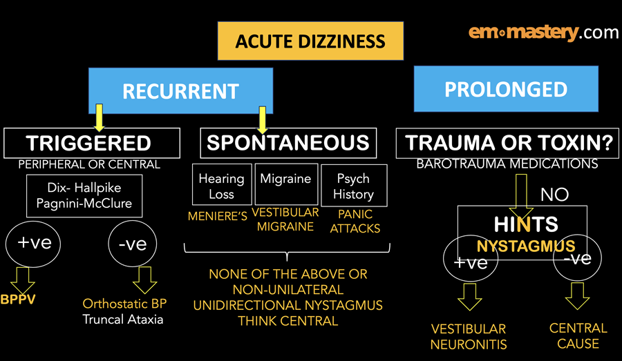
The simplified approach determines if the vertigo is acute and recurrent and then if its spontaneous or trigerred. It also looks at the prolonged causes of vertigo. The method then uses a methodical approach to the 5 minute neurological examination specifically looking at nystagmus and ataxia as well as using the Dix-Hallpike and Pagnini-McClure manoeuvres and HINTS.
The overall approach looks little the diagram below.
Maximising CPR
In this weeks video (@drpeterkas on instagram) I talk about
using POCUS to identify the best position for our hands on the chest to maximise the effectiveness of our CPR.
Everyone's heart is in a different position. Just look at the people around you. Everyone's heart cannot be in the same position, yet we push in the same place. The recommended location for compressions, is in the midline, along the inter-nipple line. It has been shown that the left ventricle is in this position in only 20% of cases.
In a retrospective study by Pickard, it was found that the best systolic blood pressure was found to be obtained by performing chest compressions to the left of the sternum, not in the lower half of the sternum as the guidelines recommendedmmend. Read the full review here.
ECG Challenge
Try these ecgs and then check the answers below.
ECG 1
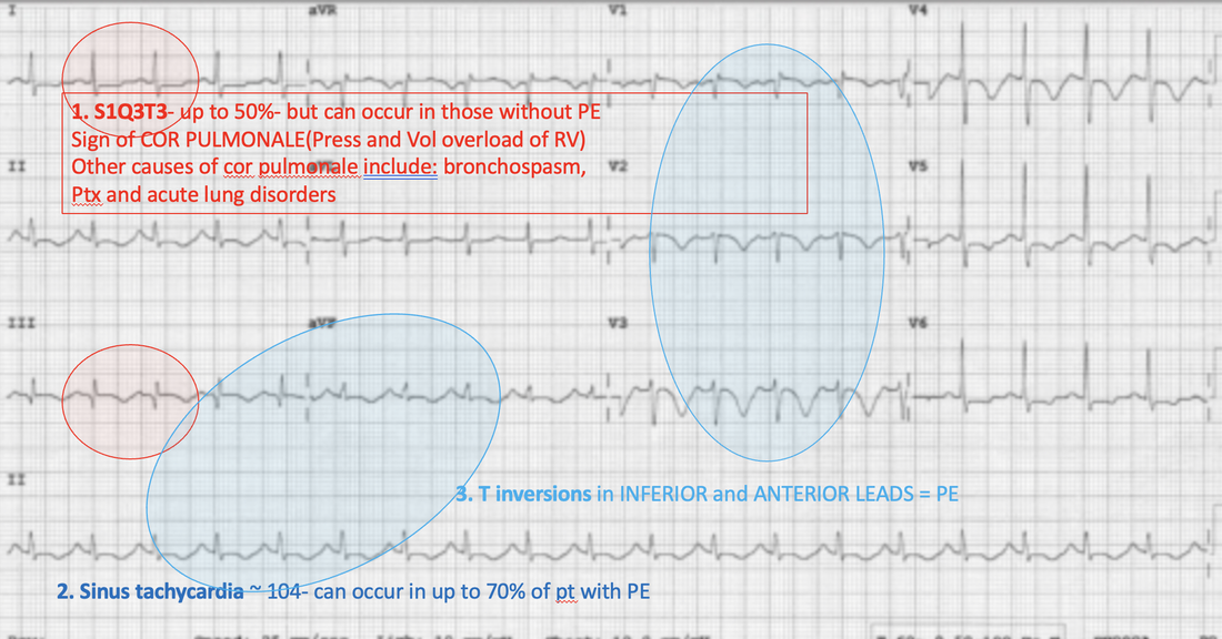
A 69 year old woman presents with dyspnoea. Her ECG is shown below. Describe the ECG and interpret the changes by giving your most likely diagnosis.

ECG 2
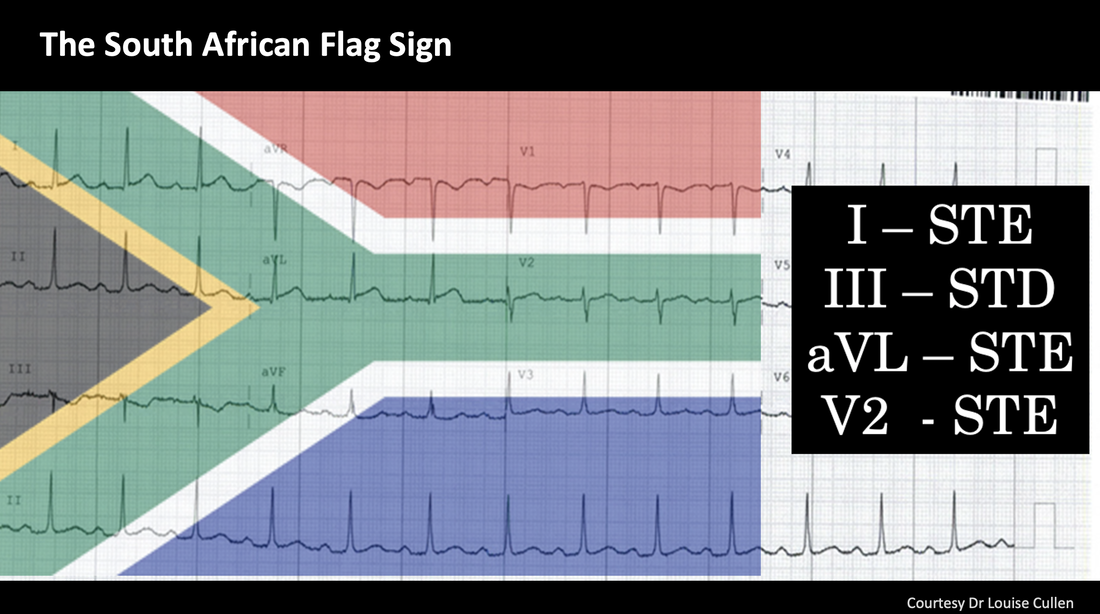
A 54 yo male presents with chest pain, that radiates into his jaw. He has a previous history of an AMI 10 years previously and also has hypertension, diabetes and hypercholesterolaemia. An ECG is done. Is this a STEMI equivalent? What is the ‘sign’ seen here?

This is a’The South African Flag Sign’. The patient had an obstructed diagonal artery.
A/Prof Peter Kas


Responses